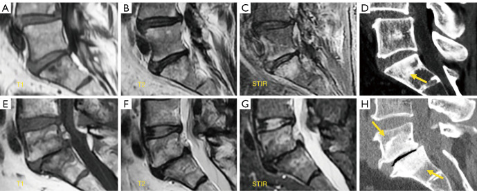
Figure 15.
Modic changes. Sagittal T1 weighted image (A), T2 weighted image (B) and STIR sequence image (C) of Modic type I changes. Sclerosis (arrow) detected at S1 on CT (D) is barely visible on MRI. Sagittal T1 weighted image (E), T2 weighted image (F) and STIR image sequence (G) of Modic type II changes. Sclerosis (arrows) conspicuous on CT (H) is barely suspected on MRI. STIR, short tau inversion recovery; CT, computed tomography; MRI, magnetic resonance imaging.

