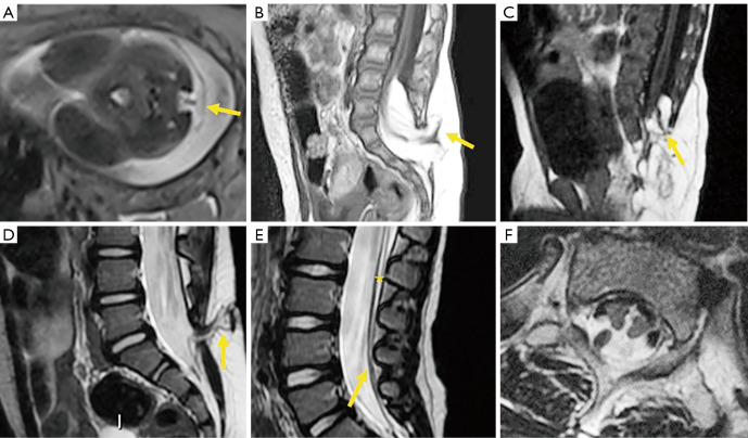
Figure 2.
Spinal dysraphism. (A) Myelomeningocele in open dysraphism in intrauterine fetus showing the protruded placode (arrow). (B) Posterior closed dysraphism with lipomyelomeningocele. The lipoma/placode interface (arrow) is outside the spinal canal. (C) Lipomyelocele The lipoma/placode interface (arrow) is within the spinal canal. (D) Dermal sinus (arrow). (E) Tethered cord with filum terminale lipoma (*) and filar thickening (arrow). (F) Split cord/diastematomyelia.

