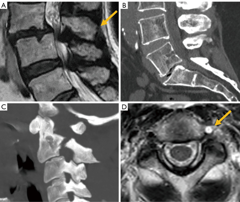
Figure 23.
Alignment abnormalities in three patients. (A) Sagittal T2 weighted image of a patient with degenerative spondylolisthesis with narrowing of the central canal and anterior shifting of the spinous process (arrow). (B) Sagittal CT with retrolisthesis of L5. (C) CT with MIP reconstruction in Hangman’s fracture. (D) Axial T2 of a hyperintense vertebral artery (arrow) secondary to arterial dissection in Hangman’s fracture. CT, computed tomography; MIP, maximum intensity projection.

