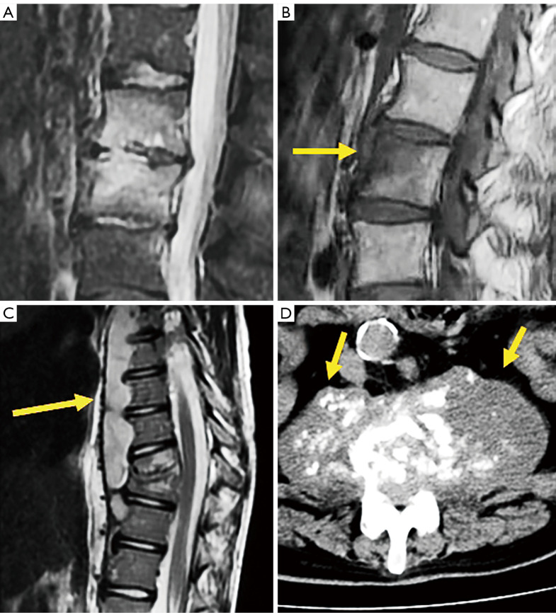
Figure 27.
Infectious pathology of the spine. (A) Sagittal STIR image of a case of pseudomonas discitis. (B) Sagittal T1 weighted image of a case of brucella osteomyelitis (arrow). (C) Sagittal T2 weighted image of a case of tuberculous prevertebral subligamentous abscess (arrow). (D) Axial CT of a case of chronic bilateral psoas abscess showing muscle enlargement with fluid collection and small calcifications (arrows). STIR, short tau inversion recovery; CT, computed tomography.

