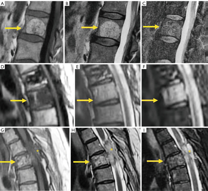
Figure 28.
Vertebral hemangioma. Sagittal T1 weighted image (A), T2 weighted image (B), and STIR sequence image (C) of typical hemangioma (arrows), hyperintense on T1 and T2 due to fat content, remaining hyperintense on STIR only the vascular content. Sagittal T1 weighted image (D), T2 weighted image (E), and STIR image sequence (F) of atypical hemangioma (arrows), hypointense on T1 due to predominance of vascular component and scarce fat content. Sagittal T1 weighted image (G), T2 weighted image (H), and STIR image sequence (I) of aggressive hemangioma (arrows) extending to the spinal canal (*). STIR, short tau inversion recovery.

