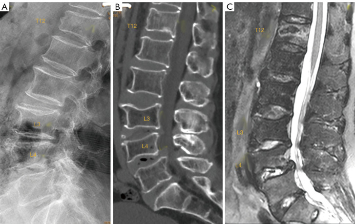
Figure 8.
Elderly patient with spine trauma. (A) Radiograph; (B) sagittally reconstructed CT; (C) sagittal T2 weighted fat suppressed MRI. A T12 traumatic fracture, L3 chronic osteoporotic upper endplate fracture, and L4 chronic osteoporotic deformity (i.e., fracture) are detected. For T12, on radiograph, attention should be paid to the anterior cortex fracture and vertebral height loss, while on MRI apparent abnormal high signal is noted. Conversely, L3 and L4 show deformity, but no abnormal high signal is noted on MRI. CT, computed tomography; MRI, magnetic resonance imaging.

