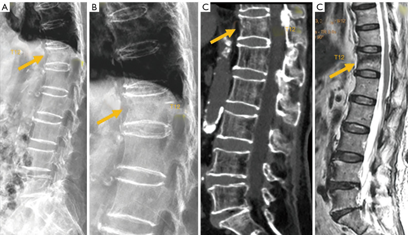
Figure 9.
Subtle osteoporotic fracture after low energy trauma (arrow). (A) Radiograph; (B) radiograph magnified view for T12; (C) sagittally reconstructed CT; (D) T1 weighted MRI. On radiograph T12 vertebral anterior cortex buckling is noted. There is no apparent height loss of the vertebral body. CT confirms T12 fracture and vertebral anterior cortex break. MRI also confirms T12 fracture. CT, computed tomography; MRI, magnetic resonance imaging.

