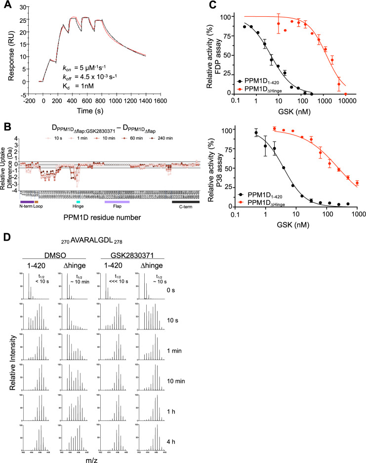
Fig. 6. The Hinge, not the flap, mediates GSK2830371 binding.
A Surface plasmon resonance of PPM1DΔflap and GSK2830371 (red represents raw data and black represents fitted kinetic parameters). B Relative differences in deuterium uptake between PPM1DΔflap in the presence of GSK2830371 compared to PPM1DΔflap in the absence of GSK2830371 as assessed by HDX-MS. Vertical tick marks between peptides on the X-axis are shown to indicate regions of missing coverage. The bars below the X-axis represent the N-terminus (purple), loop (brown), hinge (cyan), flap (light purple), and C-terminus (black) of the protein. C Activity of GSK2830371 in the FDP (top) and p38 MAPK (bottom) enzymatic assays with PPM1D1–420 (black) or PPM1DΔhinge (red). N = 2 independent samples runs. Data are presented as mean values ± SD. D EX1 analysis showing the change in deuterium uptake for peptide 270 AVARALGDL 278 located in the flap region in PPM1D1–420 and PPM1DΔhinge in the absence (left) and presence (right) of GSK2830731. Source data are provided in the Source Data file.

