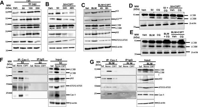
Figure 5.
CSP7 hinders silica- and BLM-induced autophagy in hAECs in vitro. (A) Representative immunoblot images showing concentration dependent expression of p53, p53S15P, LC3BII, p62 and ATG12-ATG5 in hAECs exposed silica for 72 h. (B) Representative immunoblot showing decrease in p53, p53S15P, LC3BII, p62 and ATG12-ATG5 expression in hAECs exposed to silica and treated with CSP7 for 24 and 48 h. (C) Representative Western blot images showing inhibition of p53, p53S15P, LC3BII, p62 and ATG12-ATG5 in hAECs exposed to BLM followed by CSP7 for 4 and 8 h. Representative immunoblot images showing autophagy flux analysis in hAECs (D) silica and (E) BLM exposure injury. Representative Western blot images showing immunoprecipitation (IP) of LC3B and p62 by Cav1 in hAECs exposed to (F) silica and (G) BLM, which is abrogated by CSP7 treatment. Immunoblots of IP with non-specific IgG and inputs of the corresponding lysates of hAECs serve as controls. β-actin serves as loading control. MW: Molecular weight. kDa: Kilodalton. Each experiment was repeated at least 2 times with similar results.

