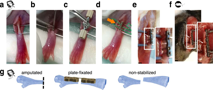
Fig. 2. Overview of the surgical procedure and the experimental setup.
a ≥20 cm snout-to-tailtip axolotl femurs were exposed by dissecting upper hind limb skin and spreading the muscles above the femur. b A titanium plate (7.75 mm Plate, RISystem, Switzerland) was attached to the femur by 4 titanium screws. c A saw guide was used for osteotomy. d A Gigly wire (0.66 mm) was inserted under the bone and used for sawing. Green outline and arrow show the plastic foil used for soft tissue protection during the surgery. e Osteotomy resulted in 0.7 mm bone gap in axolotl (white arrow). f 0.7 mm fracture of mouse femur is shown (white arrow). Here, a 6-hole plate (10 mm MouseFix Plate XL, RISystem) was attached to the femur by 4 titanium screws to fixate the bone. Scale bars 1 mm. g Sketch of the three different osteotomy models used. Amputation of the limb at the mid-femur (left) leads to blastema formation in the region of the proximal femur end of the osteotomy gap of the plate-fixated fracture (middle) and the non-stabilized fracture (right). In the non-stabilized fracture the two bone ends do not stay aligned but converge in a random way. Dashed line shows the amputation plane.

