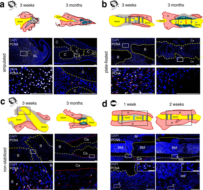
Fig. 5. PCNA immunostaining showed a decreased proliferation in amputated limbs and fractured bones at 3 months post-surgery.
Representative samples of axolotl upper hind limbs stained with anti-PCNA (white) and DAPI (blue) 3 weeks and 3 months post-surgery in axolotl amputated limbs (a), in plate-fixated fractures (b), and in fractures without fixator (c). Note high number of PCNA+ cells in 3 weeks axolotl samples. Only a few PCNA+ cells can be observed in 3 months samples. Here, proliferating cells are mostly observed in the soft tissues. d Mouse fracture samples show many PCNA+ cells in the proximity to injury site at 1 week post-fracture. At 2 weeks post-fracture, PCNA+ cells are retained in the bone marrow, and not in the newly formed bone. d’ and d” show enlarged boxed areas. Scale bars 200 µm, 50 µm in d’ and d”. Yellow dashed line - bone, red dashed line - callus, red line on sketches – former fracture site, BM – bone marrow, M – muscle, C – cartilage, Ca – callus, BL – blastema, B – bone, red arrows show PCNA+ cells.

