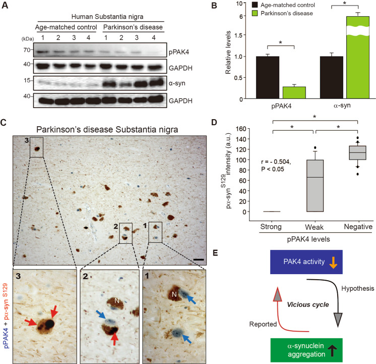
Fig. 1. Inverse correlations between pPAK4 levels and α-synuclein aggregation in the substantia nigra of postmortem brain tissue from patients with PD.
A Immunoblotting of SN lysates for pPAK4 and α-synuclein. B Quantification of the blot in A normalized to GAPDH. Age-matched controls and PD patients (n = 4 for each group). C Costaining for pPAK4 and pα-synS129. Scale bars, 25 µm. Boxed areas in C are shown at higher magnification. N neuromelanin. pPAK4, blue arrow; α-synuclein, red arrow. D Spearman correlation analysis for an inverse correlation between pPAK4 and pα-synS129 levels in human PD tissues. PD patients, n = 7; neuromelanin-positive cells, n = 63. r = −0.504, P < 0.05. E Model for the relationship between PAK4 activity and α-synuclein aggregation. The data are presented as the mean ± SEM. *P < 0.05. Unpaired Student’s t test (B) or two-way ANOVA (D) was used for statistical analysis followed by Tukey’s multiple comparisons test.

