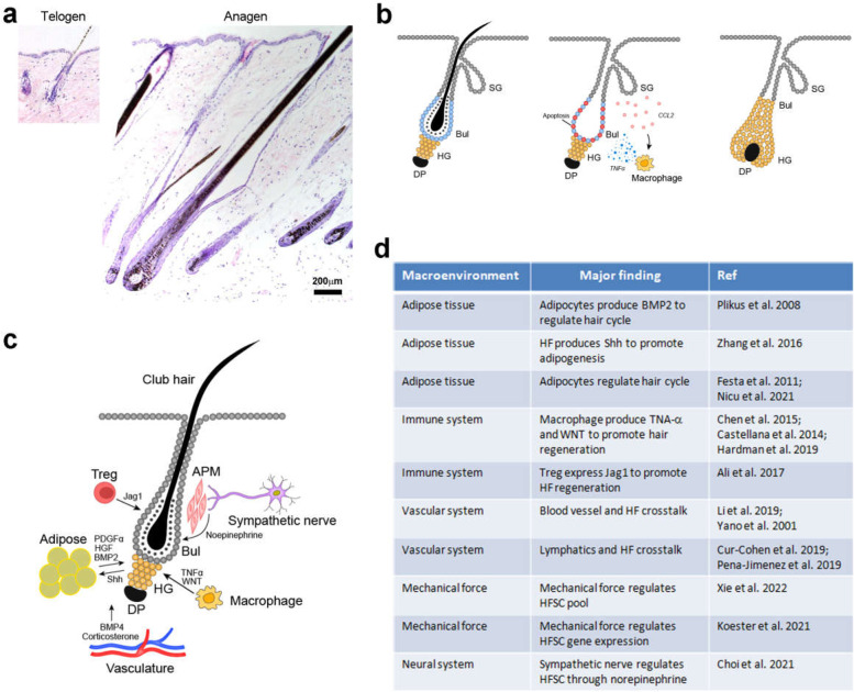
Fig. 1.
Regulation of hair regeneration. A Haematoxylin and eosin staining showing the mouse HF in resting (telogen; left) and growing (anagen; right) phases. The panels are of the same magnification to show the difference in size. B Diagrams showing the hair regeneration process. The hair germ (HG) is the population of progenitors between the dermal papilla (DP) and the bulge stem cells (Bul). Plucking induces bulge stem cell apoptosis, and Ccl2 secretion from the wound epithelium, followed by activation of macrophage which secrets TNFα to promote hair regeneration. SG, sebaceous gland. C A diagram summary of the various layers of regulation on hair regeneration. Sympathetic nerves produce norepinephrine to activate HFSCs in the bulge (Bul). Macrophages produce TNFα (in wound) and Wnt ligands (in natural telogen-anagen transition) to activate hair growth. Adipocyte precursors secret PDGFα/HGF to promote hair growth, and adipocytes produce BMP2 to help maintain telogen, whereas these cells sense SHH produced by actively growing HFs to proliferate. Treg cells express Jag1 to activate Notch signaling and HFSCs. The vasculature systems including blood vessels and lymphoid vessels are also important for hair growth and regeneration. Circulating hormones such as corticosterone act on the DP to regulate hair growth. D Summary of the various macro-environmental factors regulating HFSCs and hair regeneration

