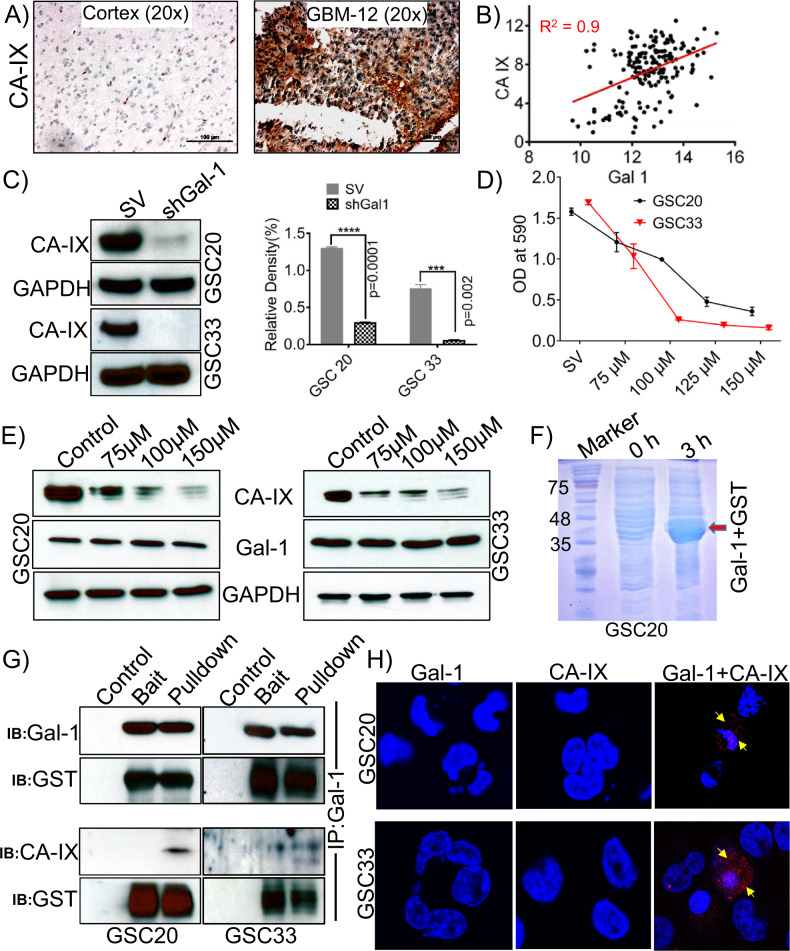
Fig. 5. Carbonic anhydrase (CA-IX) is highly expressed in GBM; Gal-1 regulates the expression of CA-IX.
A Immunohistochemical staining images of CA-IX expression in control brain and GBM specimen (Scale bar-100 μm). B The mRNA obtained for Gal-1 and CA-IX from TCGA datasets showed a positive correlation. C Western blot analysis of CA-IX in shGal-1 stable and SV treated GSC20 and GSC33 cells. The density levels were quantified and represented as a bar graph. D MTT assay. E Expression of CA-IX and Gal-1 in untreated controls and samples treated with different concentrations of CA-IX inhibitor, SLC-0111, in GSC20 and GSC33 cells. F Coomassie Blue-stained SDS-PAGE gel showing the expression levels of GST-Gal-1 protein after induction with 0.5 mM IPTG for 3 h. G Co-immunoprecipitation (GST pulldown) assay was performed with Gal-1 and CA-IX antibodies, using GST-tagged Gal-1 fusion purified protein lysate as prey and GSC20/GSC33 control total protein as bait. Eluted proteins were resolved by electrophoresis and subjected to immunoblot analysis. GST is shown as a loading control. H PLA of Gal-1 and CA-IX were performed in GSC20 and GSC33 cells. The representative micrograph of Gal-1 + CA-IX shows the positive PLA. Red spots confirmed the association. Single antibody stain with Gal-1 and CA-IX showed negative results. DAPI was used to stain the nuclei.

