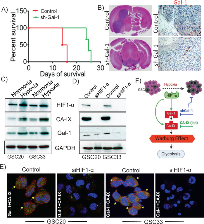
Fig. 6. Silencing Gal-1 suppresses the growth of GSC33 generated tumor in athymic nude mice.
Approximately 50,000 SV-treated and GSC33 shGal-1 cells were intracranially implanted into the brain of 4-week-old male and female athymic nude mice using stereotaxic apparatus (n = 8). A Kaplan–Meier curves plotted to show a significant increase in percent survival for all shGal-1-treated mice (n = 8) (p < 0.05) compared to untreated tumor-bearing mice (n = 8). B Hematoxylin and Eosin staining confirmed the survival data. Immunohistochemistry analysis performed on untreated and shGal-1-treated mice showed significant reduction in the expression of Gal-1 (Bar = 100 µm). C Immunoblot analysis shows the expression profiles of HIF-1α, Gal-1, and CA-IX in normoxia and hypoxia conditions. D GSC20 and GSC33 cells were transfected with siRNA against HIF-1α and the indicated proteins were analyzed by immunoblot analysis. E The siHIF-1α transfected GSC20 and GSC33 cells were labeled with Gal-1 and CA-IX antibodies. Immunocytochemistry shows increased colocalization of Gal-1 and CA-IX in both the controls whereas the siHIF-1α cells showed a reduced expression (Red = Gal-1; Green = CA-IX; Blue = DAPI; colocalization = Yellow). E Schematic representation of the possible regulation of HIF-1α-Gal-1-CA-IX axis in promoting Warburg effect in GSC.

