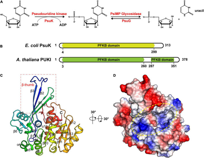
FIGURE 1.
Structure of apo-EcPsuK. (A) Reaction scheme of pseudouridine metabolism. (B) Domain organization of Arabidopsis PUKI and E. coli PsuK. (C) Overall structure of EcPsuK is colored in the rainbow. β-Thumb region is indicated by a red box. (D) Surface representation of apo-EcPsuK, and the potential substrate-binding pocket is indicated by a yellow oval.

