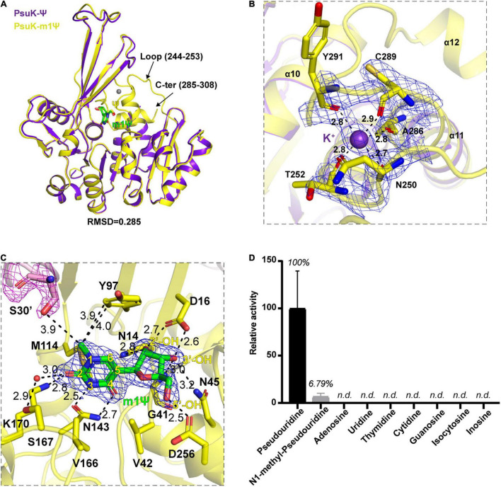
FIGURE 5.
Structure complex of EcPsuK with m1Ψ. (A) Structure superposition of EcPsuK-m1Ψ and EcPsuK-Ψ. m1Ψ is colored in green, and Ψ is colored in yellow. The lost regions in the EcPsuK-Ψ complex are indicated by black arrows. (B) Binding site of a monovalent ion. The 2| Fo| –| Fc| σ-weighted map is contoured at 1.5σ. K+ is colored purple shown as a sphere. (C) Detailed interactions between EcPsuK and m1Ψ. EcPsuK is shown as cartoon and colored in yellow, the residues in contact with Ψ are shown as sticks, and m1Ψ is shown as stick colored in green. The hydrogen bonds are shown as black dashed lines. The red sphere represents the water molecule. The 2| Fo| –| Fc| σ-weighted map is contoured at 1.5σ. (D) Substrate specificity of EcPsuK for pseudouridine and other nucleoside analogs. n.d., not detected.

