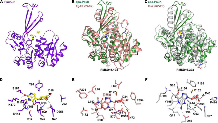
FIGURE 6.
Comparisons of substrate binding properties of EcPsuK with TgAK and Gsk. (A) The overall structure of EcPsuK in complex with Ψ. (B) Structure superposition of EcPsuK-Ψ with TgAK-DMA complex (PDB code: 2A9Y). The latter complex structure is colored in pink. (C) Structure superposition of EcPsuK-Ψ with Gsk-guanosine complex (PDB code: 6VWP). The latter complex structure is colored in white. (D) Detailed interactions between PusK with Ψ. (E) Detailed interactions between TgAK with DMA (N6, N6-dimethyladenosine). (F) Detailed interactions between Gsk with guanosine.

