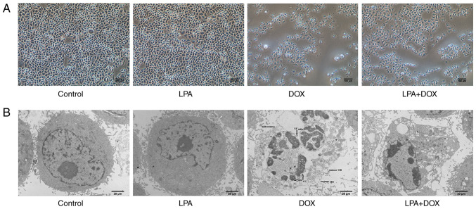
Figure 1.
Morphology of HeLa cells treated with DOX and LPA. (A) Images were captured using an optical microscope. Magnification, ×100. Scale bar, 100 µm. (B) Images were captured using a transmission electron microscope. Magnification, ×2,000. Scale bar, 20 µm. Arrows indicate membrane broken, nuclear fragmentation, and chromatin condensation. DOX, doxorubicin hydrochloride; LPA, lysophosphatidic acid; MB, membrane broken, NF, nuclear fragmentation; CC, chromatin condensation.

