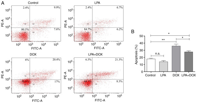
Figure 2.
Determination of the apoptotic rate using flow cytometry. (A) Flow cytometry was used to detect apoptosis in the control, LPA, DOX and LPA + DOX HeLa cell groups. The percentage of apoptotic cells in Q1, Q2 and Q4 are as indicated. (B) Quantification of the apoptotic rate was determined using the flow cytometric data. Data are presented as the mean ± SD (n=3). *P<0.05 and **P<0.01 vs. the control. DOX, doxorubicin hydrochloride; LPA, lysophosphatidic acid; n.s., no significance.

