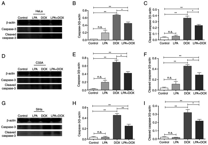
Figure 4.
Effect of LPA on the protein expression levels of caspase-3 and cleaved caspase-3 in DOX-induced HeLa, C33A and SiHa cells. After HeLa, C33A and SiHa cells were treated as the control or with LPA, DOX or LPA + DOX for 24 h, the cells were harvested and lysed for western blotting. (A) Western blotting was performed to determine caspase-3 and cleaved caspase-3 protein expression levels in HeLa cells. (B) Semi-quantification of caspase-3 protein expression levels in HeLa cells was performed using ImageJ software. (C) Semi-quantification of cleaved caspase-3 protein expression levels in HeLa cells was performed using ImageJ software. (D) Western blotting was performed to determine caspase-3 and cleaved caspase-3 protein expression levels in C33A cells. (E) Semi-quantification of caspase-3 protein expression levels in C33A was performed using ImageJ software. (F) Semi-quantification of cleaved caspase-3 protein expression levels in C33A cells was performed using ImageJ software. (G) Western blotting was performed to determine caspase-3 and cleaved caspase-3 protein expression levels in SiHa cells. (H) Semi-quantification of caspase-3 protein expression levels in SiHa cells was performed using ImageJ software. (I) Semi-quantification of cleaved caspase-3 protein expression levels in SiHa cells was performed using ImageJ software. Data are presented as the mean ± SD (n=3). *P<0.05 and **P<0.01 vs. the control. DOX, doxorubicin hydrochloride; LPA, lysophosphatidic acid; n.s., no significance.

