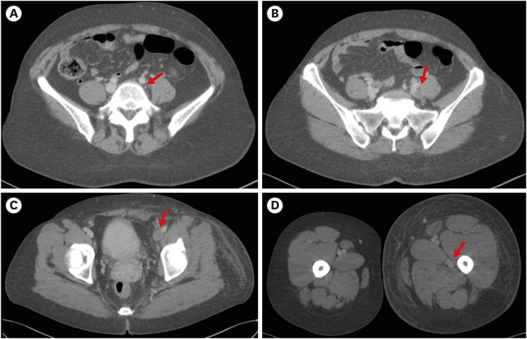
Fig. 2. Initial CT of lower extremity. CT scan shows that the left iliac vein is externally compressed (arrow) between the right common iliac artery and spinal body (A), resulting in complete occlusion of the left iliac vein (B) with extensive thrombus (arrows) involving the left iliac vein (B), left femoral vein (C), and left popliteal vein (D). The patient’s left lower extremity exhibits marked swelling.
(B, C, D) Arrows indicate thrombus in the left iliac, left femoral, and left popliteal vein, respectively.
CT = computed tomography.

