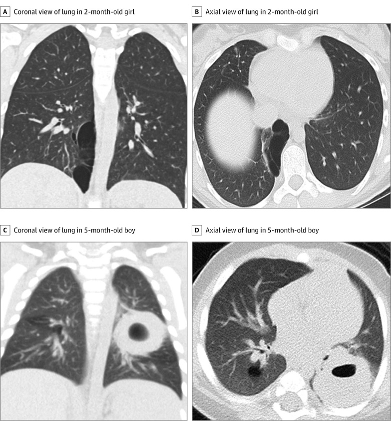
Figure 2. Examples of Computed Tomography Images Reviewed by Study Radiologists.
Representative coronal (A) and axial (B) lung window images in a 2-month-old girl show a 5.5 × 2.6 × 8.9-cm cystic lung lesion located in the medial right lower lobe with thin internal septations. The pathologic diagnosis was pleuropulmonary blastoma (PPB) but was interpreted by 6 study radiologists as a benign macrocystic congenital pulmonary airway malformation (CPAM). Representative coronal (C) and axial (D) lung window images in a 5-month-old boy with a posterior left lower lobe 2.9 × 3.5 × 2.7-cm solid lung lesion with an internal cyst. The pathologic diagnosis was CPAM but was diagnosed by 6 study radiologists as PPB.

