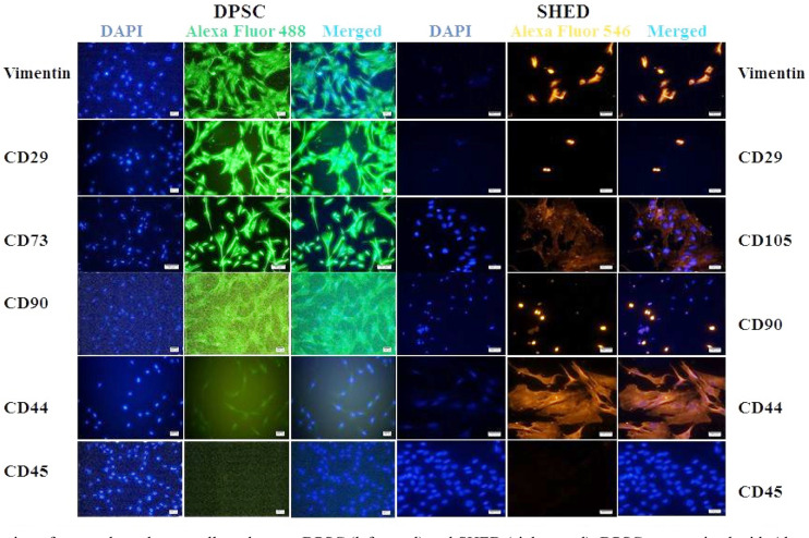
Fig.3.
Expression of mesenchymal stem cell markers on DPSC (left panel) and SHED (right panel). DPSC stained with Alexa Fluor 488 (green) show the expression of vimentin, CD29, CD73, CD90, and CD44 in DPSC. SHED were stained with Alexa Fluor 546 (orange) show the expression of vimentin, CD29, CD105, CD90, and CD44. CD45 was used as negative marker. Micrographs were captured with Ti2 inverted microscope and analyzed using NIS element software. Scale bar= 100 μm and 50 μm for CD73 images only.

