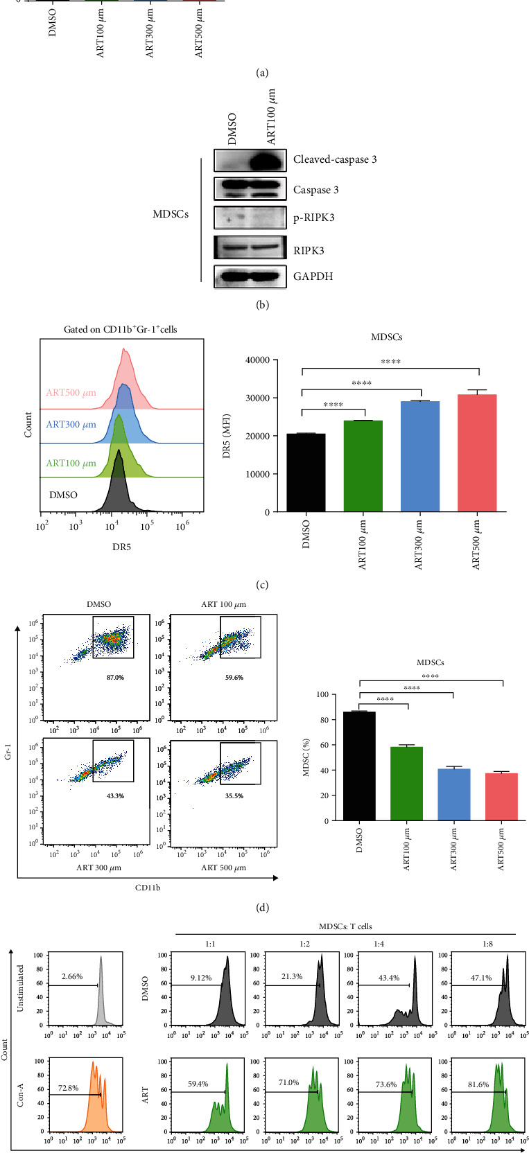
Figure 1.

ART promotes MDSC apoptosis and inhibits the accumulation and immunosuppressive function of MDSCs. (a–e) Bone marrow (BM) cells isolated from wild-type C57BL/6 mice were cultured with GM-CSF and IL-6 for 3 days to generate in vitro BM-derived MDSCs and then were treated with different concentrations of ART (100 μM, 300 μM, and 500 μM) for another 12 hours, and the solvent DMSO was used as the control. (a) The apoptosis levels of CD11b+Gr-1+ MDSCs were detected by flow cytometrical analysis. (b) The cleaved-caspase3, caspase3, p-RIPK3, and RIPK3 of CD11b+ Gr-1+ MDSCs were detected by western blotting. (c) The DR5 mean fluorescence intensity of CD11b+ Gr-1+ MDSCs was detected by flow cytometrical analysis. (d) The proportion of CD11b+ Gr-1+ MDSCs was detected by flow cytometric analysis. (e) Flow cytometry to purify CD11b+Gr-1+ cells from in vitro 100 μM ART-treated BM-derived MDSCs and further cocultured CD11b+Gr-1+ MDSCs with Con A-stimulated CD3+ T cells isolated from the spleens of wild-type C57BL/6 at the 1 : 1, 1 : 2, 1 : 4, and 1 : 8 ratios to detect the percentages of proliferation T cells as tested by CFSE fluorescence. Data are means ± SEM and are from a representative experiment of three (a–e) independent experiments. Unpaired Student's t test for (a)–(e). ∗P < 0.05, ∗∗∗P < 0.001, and ∗∗∗∗P < 0.0001.
