
Figure 5.
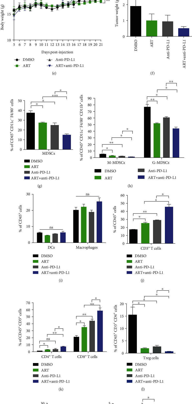
Targeting MDSCs via ART therapy significantly enhances the efficacy of anti-PD-L1 immunotherapy in tumor-bearing mice. (a–o) In B16F10 or Hepa 1-6-bearing mice, we treated with 50 mg/kg ART daily while administrated 10 mg/kg anti-PD-L1 antibodies once every three days, starting from days 9 and 5 following injection of B16F10 or Hepa 1-6 cells, respectively (n = 5 mice per group). (a and d) Tumor growth curves, (b and e) mice weight growth curves, and (c and f) tumor weights recorded from B16F10 or Hepa 1-6 tumor-bearing mice. (g–n) The proportions of immune cells in tumor tissues of Hepa 1-6 tumor-bearing mice: MDSCs, M-MDSCs, G-MDSCs, DCs, Macrophages, CD3+ T cells, CD4+ T cells, CD8+ T cells, Treg cells, B cells, and NK cells were detected by flow cytometry. (o) Tumor MDSCs isolated from Hepa 1-6-bearing mice were treated with the combination therapy of ART and anti-PD-L1 antibodies cocultured at 1 : 2 ratio with CFSE-labeled spleen CD3+ T cells purified from wild-type C57BL/6 mice activated with Con A (5 μg/ml). The cells were cultured for 3 days and stained with CD3 antibody, and T cell proliferation was analyzed by flow cytometry. Data are means ± SEM and are from a representative experiment of three (a–n) or from two (o) independent experiments. Unpaired Student's t test for (a)–(n). ∗P < 0.05, ∗∗P < 0.01, ∗∗∗P < 0.001, and ∗∗∗∗P < 0.0001. ns: not significant.