. 2022 Jul 3;19(4):445–455. doi: 10.21873/cgp.20331

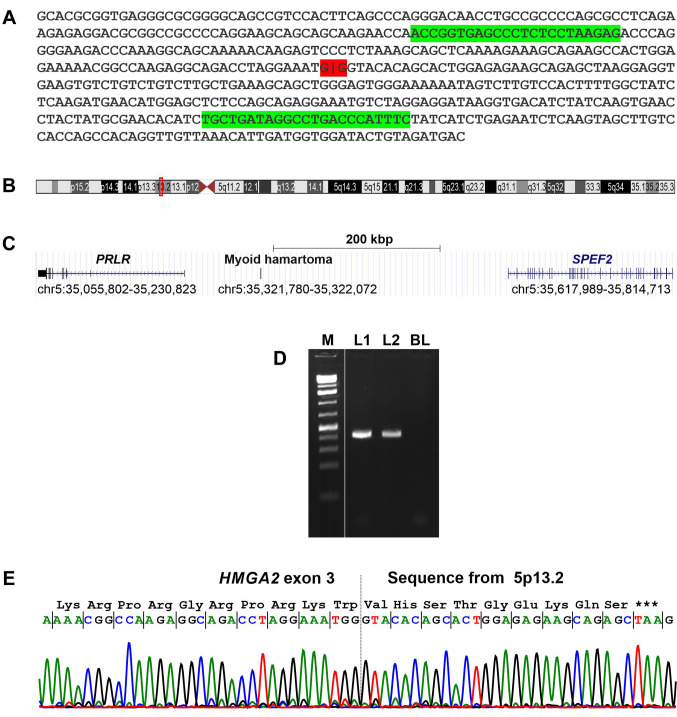
Figure 3. Molecular genetic examination of the mammary leiomyomatous tumor. A: The high-mobility group AT-hook 2 (HMGA2) fusion sequence was obtained from raw RNA sequencing data using the deFuse software package. The G|G junction of HMGA2 with a sequence from chromosome sub-band 5p13.2 is highlighted in red. The position of the forward HMGA2-929F1 and reverse 5p13R primers are highlighted in green. B: Ideogram of chromosome 5 showing the position of the sequence of myoid hamartoma found by RNA sequencing/deFuse software to have fused with exon 3 of HMGA2, together with the prolactin receptor (PRLR) and sperm flagellar 2 (SPEF2) genes in sub-band 5p13.2 (red box). C: Diagram showing the 768-kbp region of the sub-band 5p13.2 which contained the sequence of myoid hamartoma, PRLR, and SPEF2 together with their genomic positions on the GRCh37/hg19 assembly. D: Gel electrophoresis showing the amplified cDNA fragment using the forward primer HMGA2-929F1 and the reverse 5p13R. M: 1-kb DNA ladder (GeneRuler, ThermoFisher Scientific). L1: The fragment from the positive control (previously published myoid hamartoma). L2: The fragment from the present myoid hamartoma of the breast. BL: Blank, water as template in the polymerase chain reaction. E: Partial sequence chromatograms of the cDNA amplified fragment showing the junction position of HMGA2 and the sequence from chromosome sub-band 5p13.2 (vertical dotted line) together with part of the coding peptide. ***Denotes stop of translation.

Figure 3