. 2022 Jul 3;19(4):415–427. doi: 10.21873/cgp.20329

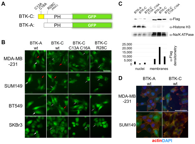
Figure 3. Subcellular distribution of the BTK isoforms in breast cancer cells. (A) Diagram of BTK pleckstrin homology domain with GFP reporters. Expression of each reporter was driven by a CMV promoter in a retroviral vector. (B) Localization of wild type and mutant isoform reporters in breast cancer cell lines MD-AMB-231 (triple negative; PTEN+), BT549 (triple negative; PTEN), SUM149 (triple negative; PTEN) and SKBr3 (luminal HER2 enriched; PTEN+). BTK-A isoform is found in the plasma membrane (red arrows) and to a greater degree in the nuclei of cancer cells (white arrows). BTK-C is also found on the plasma membrane but exhibits a greater degree of perinuclear localization (gray arrows). Mutation of the two palmitoylation sites reduces membrane localization of BTK-C. Mutation of arginine 28 of BTK (arginine 62 and BTK-C sequence) also decreases membrane staining. (C) Subcellular fractionation showing increased levels of BTK-A in the nucleus and greater BTK-C in membrane fractions. (D) BTK reporters localize with actin in cancer cells. Scale bars: 100 μm.

Figure 3