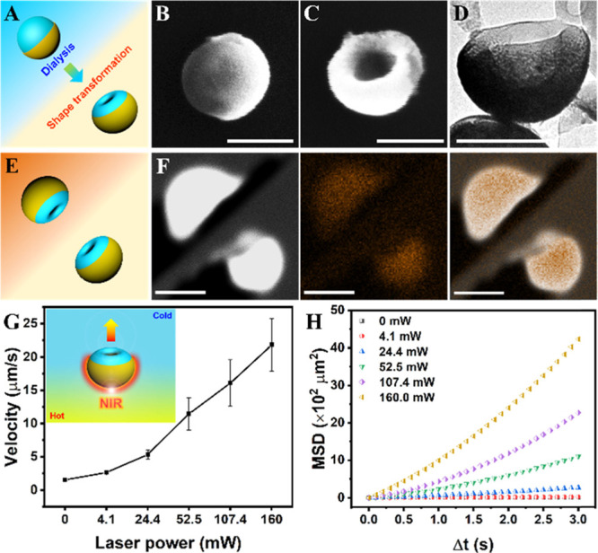
Figure 2.

Preparation and characterization of Janus stomatocyte-based supramolecular nanomotors (motor 1: Janus stomatocytes) and their NIR triggered motion. (A) Schematic illustration of the construction of Janus stomatocytes via dialysis treatment. (B) SEM image of a Janus polymersome before dialysis, scale bar = 200 nm. (C) SEM image of a Janus stomatocyte after dialysis, scale bar = 200 nm. (D) TEM image of a Janus stomatocyte, scale bar = 200 nm. (E) Schematic depiction of the orientation of Janus stomatocytes for energy-dispersive X-ray spectroscopy (EDX) elemental mapping analysis. (F) Elemental mapping of Janus stomatocytes by EDX showing the Janus morphology of stomatocytes. From left to right: electron image, EDX mapping image of Au, and merged image. Scale bar = 500 nm. (G) Velocity dependence of the Janus stomatocytes on the NIR output laser power. (H) Mean square displacement (MSD) of Janus stomatocytes versus time interval (Δt) analyzed from motion tracking trajectories.
