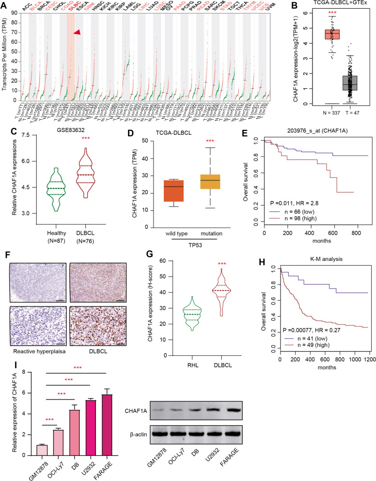
Fig. 1.
CHAF1A is highly expressed in DLBCL compared with benign healthy tissues. A Pan-cancer profiles based on GEPIA2 platform showing the expression levels of CHAF1A in multiple malignancies matched with respective normal samples, individually. B Differential analysis based on TCGA-DLBCL cohort revealing that CHAF1A was elevated in DLBCL (N = 47) versus GTEx normal tissues. C Besides, CHAF1A was up-regulated in DLBCL (N = 164) as compared to normal samples in the GSE83632 dataset. D Based on TCGA-DLBCL samples, TP53-mutated samples exhibit high CHAF1A expressions. E Prognostic analysis with Kaplan–Meier survival curves indicated that DLBCL samples with high CHAF1A shorter overall survival months relative to those with low CHAF1A expressions based on the GSE32918 data set (log-rank test p = 0.011). F As evidenced by IHC assay, CHAF1A expressions were notably higher in DLBCL tissues versus RHL. Upper scale bar = 200 μm, lower scale bar = 50 μm. G Differential test analysis of quantified CHAF1A expressions (h-score) in collected DLBCL and RHL samples (N = 70). H High CHAF1A expression in DLBCL correlated with shorter overall survival months based on the analysis of the IHC levels, as revealed by the Kaplan–Meier survival curve analysis (n = 90, p < 0.001). I The RT-qPCR analysis indicated the upregulated expression of CHAF1A in multiple DLBCL cell lines. Experiments were performed in triplicate. *p < 0.05, **p < 0.01, ***p < 0.001

