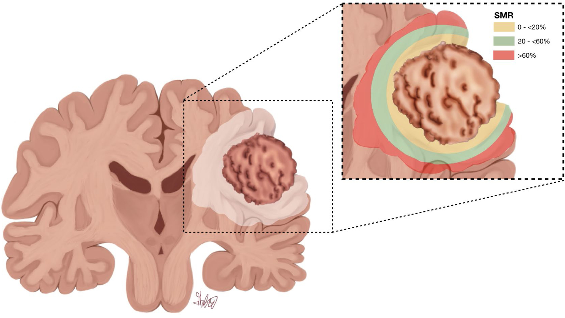
Figure 5:

SMR percentages significantly associated with beneficial OS in patients with IDH-wt GBM who underwent GTR of CE tumor. A coronal section of the brain is shown, illustrating the CE portion of the tumor surrounded by FLAIR hyperintensity, as well as a color-coded magnified view of the tumor region (inset). Yellow represents proximal SMR percentages that did not show a significant benefit in OS. Green represents SMR percentages that were significantly associated with benefit in OS. Red represents the distal FLAIR-hyperintense area in which SMR was not significantly associated with OS. Copyright Alfredo Quinones-Hinojosa. Published with permission.
