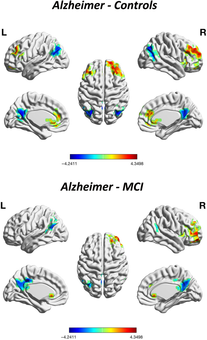
FIGURE 4.

Whole‐brain voxel‐level secondary gradient comparisons among the Alzheimer's disease (AD), mild cognitive impairment (MCI), and healthy control (HC) groups with AlphaSim correction. Blue and red clusters denote regions with significantly decreased and increased gradients, respectively (p <.05, cluster size ≥392.7 voxels, Alphasim corrected). Compared with HCs, patients with AD exhibited significant functional gradient increases mainly in the bilateral MFG, OFC, MFGtriang, and ACC, and decreases mainly in the bilateral ANG, MOG, MTG, and PCUN/PCG. Compared with patients with MCI, patients with AD exhibited a similar gradient changing pattern, showing significant increase of gradient values mainly in the MFG.R, OFC.R, MFGtriang.R, insula.R, and the bilateral ACC and decrease mainly in bilateral ANG, MTG, and PCG. There were no differences between patients with MCI and HCs in gradient values after correction
