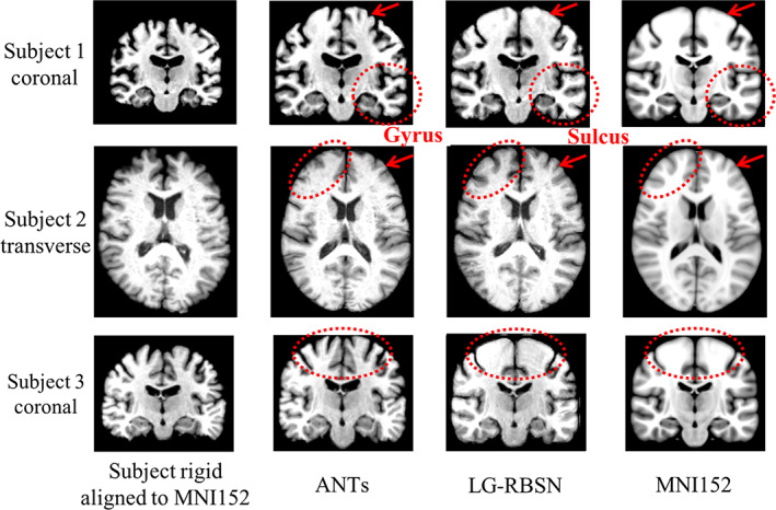
FIGURE 6.

Spatial normalized brain qualification evaluation comparison between LG‐RBSN and ANTs. The first and the last columns illustrate subjects' brain images rigid aligned (showing as moving images) to MNI152 brain images (as fixed images). The second and the third columns illustrate subjects' brain images after ANTs/LG‐RBSN nonlinear registration to MNI152 brain images. LG‐RBSN shows clearly better performance compared to ANTs in red dotted circles highlighted areas. In subject 2, ANTs mismatched a gyrus of the subject's cerebral cortex to a sulcus in MNI152 space, whereas LG‐RBSN matched the corresponding sulci properly. ANTs, advanced normalization tools; LG‐RBSN, landmark‐guided region‐based spatial normalization
