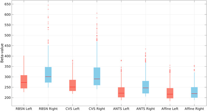
FIGURE 11.

Distribution of the point estimates for visual task‐based fMRI group level visual activation in brain contralateral occipital lobe using different spatial normalization methods (left: for stimulating left visual hemifield; right: For stimulating right visual hemifield). ANTs, advanced normalization tools; CVS, combined volumetric and surface registration; LG‐RBSN, landmark‐guided region‐based spatial normalization
