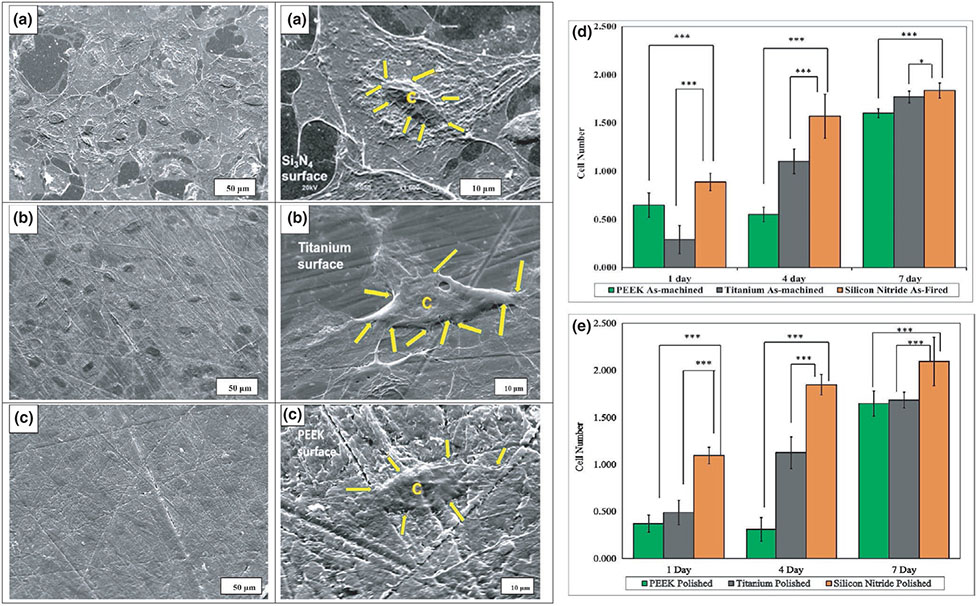
FIGURE 10.
SEM images of cell adhesion on (a) silicon nitride (as-fired), (b) Ti6Al4V (as-machined) and (c) PEEK (as-machined), C represents the cell and yellow arrows shows the filopodia on these surfaces, (d) and (e) represent cell proliferation (MTS assay) with MC3T3-E1 cells; (d) shows as-machined PEEK, Ti6Al4V and as-fired Si3N4, (e) shows polished samples of PEEK, Ti6Al4V and Si3N4

