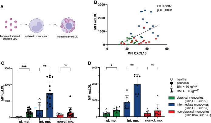
Figure 2.
Phagocytosis of oxLDL by monocytes increases with CXCL16 expression. (A) PBMC were incubated with red fluorescing oxLDL for 3 h and subsequently stained for CD14, CD16 and CXCL16 expression. (B) The mean fluorescence intensity (MFI) of CXCL16 and oxLDL is shown. Every psoriasis patient is represented by three dots (n= 15). Colors represent monocyte subsets as defined in Figure 1A , Spearman correlation. (C) oxLDL concentrations in monocytes subsets of healthy controls (unfilled dots; n=5) and patients with psoriasis (filled dots; n=15), mean and SD, unpaired t test. (D) intracellular oxLDL concentrations in monocytes of normal-weight psoriatic patients (unfilled triangles; n=7) and obese patients (filled triangles; n=8), mean and SD, unpaired t test. *p < 0.05; **p < 0.01; ***p < 0.001. ns, not significant.

