Figure 2.
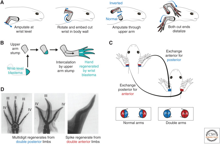
Properties of positional memory in the salamander limb. (A) Butler's circularization surgery to generate distoproximal inverted limbs. The limb is amputated at the wrist level, then the exposed surface embedded into a pocket made in the body wall muscles posterior to the shoulder. After healing and innervation, an amputation through the upper limb releases two arm stumps: one with normal proximodistal polarity (bottom arm) and the other with inverted, distoproximal polarity (top arm). Both arm stumps distalize and regenerate upper arm, lower arm, and hand elements in sequence. This experiment showed that salamander cells are constrained to generating distal, but not proximal, cells during regeneration (rule of distal transformation). The ulna (posterior lower arm bone) and posterior wrist and digit elements are shaded in red to denote the anteroposterior axis. Note that the inverted arm regenerates a mirror image limb compared with the normal arm (compare red shading). (Schematics after Dent 1954.) (B) The rule of distal transformation as shown through an intercalation assay. Transplantation of a distal, wrist-level blastema (turquoise) to a proximal, upper arm stump (gray) results in a normally patterned regenerate. The wrist-level blastema regenerates only wrist and hand, whereas the upper arm stump intercalates the intervening arm elements. (C) Generation of surgical “double” arms in which specific positional memories are deleted. Double anterior arms are generated by removing the posterior half (blue) of a recipient arm and grafting in its place the anterior half (red) from a donor arm. Fusion along the midline generates an arm lacking posterior identity. Similar operations can be used to generate double posterior arms, and also double dorsal or double ventral arms (not shown). Skeletal elements were not transplanted in this assay. (Schematic after Tank 1978.) (D) Double posterior limbs amputated immediately after construction in axolotls regenerated multidigit limbs (left and center). Note that these regenerates are often symmetrical about the midline and can harbor more digits than normal (four-digit) limbs. In contrast, double anterior limbs often failed to regenerate, or regenerated hypomorphic “spike” structures (right). (Images in Panel D from Holder et al. 1980; reprinted, with permission, from Elsevier © 1980.)

