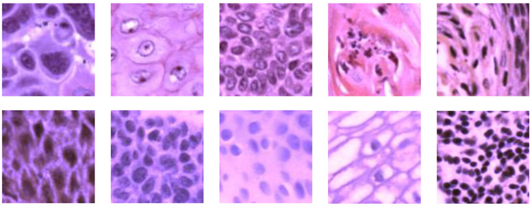
Figure 3.
Pseudo-RGB histological patches (101 × 101 pixels) showing anatomical diversity. First row: Patches extracted from cancerous ROIs with various histological features. Second row: Patches generated from images of normal ROIs, showing features at different layers of the stratified epithelium as well as the region under epithelium with inflammation.

