Figure 2.
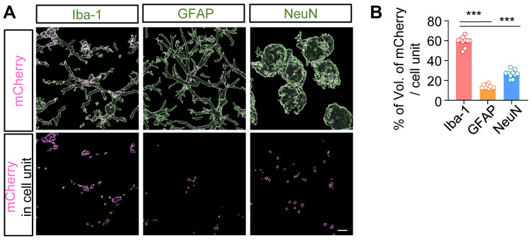
AAV-EF1α-mCherry-PLGA NPs were i.t. administered to cisterna magna: (A) On day 3 after injection, tissue of brain was isolated and used for immunostaining with anti-Iba-1, GFAP, and NeuN antibodies. The images of red fluorescence were obtained only with the expression of mCherry observed in each cell type. (B) The volume of mCherry fluorescence was quantified using the IMARIS program.
Notes: (B) Data are presented as the mean ± SEM, analyzed by one-way ANOVA followed by Tukey’s post hoc test, ***P < 0.01 compared with the other cell type.
Abbreviations: AAV, adenovirus vector; PLGA, poly lactic-co-glycolic acid; NP, nanoparticle; i.t., intrathecally; GFAP, glial fibrillary acidic protein.

