Figure 4.
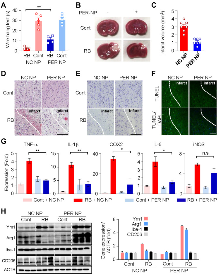
Microglia targeted by PER encapsulated in PLGA NPs attenuate infarct damage in the RB model, and reduce production of pro-inflammatory cytokines: (A) Wire hang test evaluated 24 h after RB photothrombosis. (B) Representative images of coronal brain sections stained with TTC. (C) Quantitative analysis of infarction volume. (D) Hematoxylin and eosin (H&E) staining of brain tissue sections of rat models of cerebral ischemia. Scale bar: 100 nm. (E) Cresyl violet staining of brain sections showed the infarct volume after 1 days of Rose Bengal modeling. Scale bar: 100 nm (F) Representative images of TUNEL staining in the cortical core infarct and penumbra within tissue collected after 24 h of Rose Bengal photothrombosis. Scale bar: 200 nm (G) The mRNA levels of TNF-α, IL-1β, Cox2, IL-6, and iNOS were measured using qRT-PCR. (H) Western blot analysis of Ym1, Arg1, Iba-1, CD206, and ACTB levels in cortical tissue of hemisphere subjected to 24 h of RB-induced photothrombosis.
Notes: (A) Data are expressed as mean ± SEM. **P = 0.0017 for RB/NC NP group vs RB/PER NP group. (D) On day 1 after RB modeling, total mRNA was extracted from the brain infarct region (0.7 cm) and used for cDNA synthesis. Data are presented as mean ± SEM. In one-way ANOVA with Tukey’s post hoc test, **P = 0.0016, *P = 0.0130, n.s., not significant. (G) The integrated optical density of the protein band was calibrated relative to ACTB. Data are expressed as mean ± SEM.
Abbreviations: PER, perampanel; PLGA, poly lactic-co-glycolic acid; NP, nanoparticle; RB, Rose Bengal; TTC, Triphenyltetrazolium chloride; SEM, standard error of mean; ACTB, actin beta; NC, negative control.

