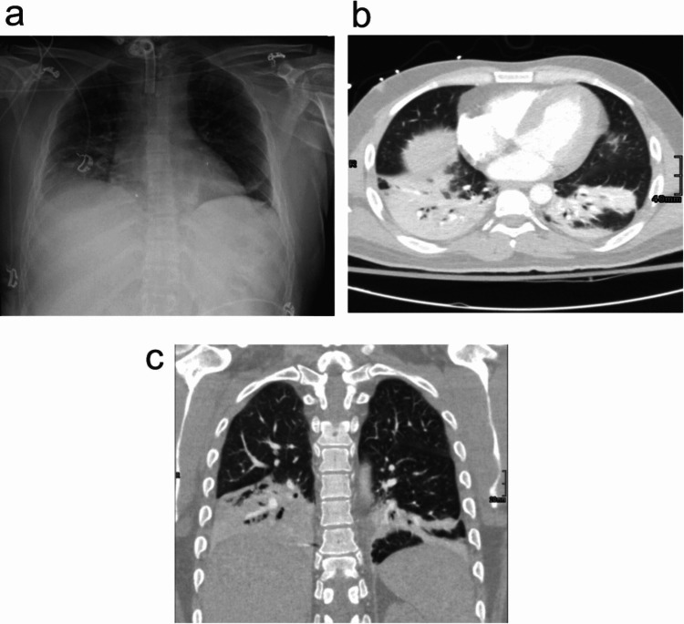
Figure 2. Radiologic findings in patients with nervous system injuries and developing pneumonia.
a. The radiograph demonstrates right basilar pleuroparenchymal and left basilar linear airspace opacities. No large pleural effusion or pneumothorax is identified.
b. Axial view of the CT chest with contrast which was performed the next day for evaluation of a possible pulmonary embolism shows bibasilar consolidative opacities with air bronchograms. No segmental or sub-segmental pulmonary embolism was identified.
c. Coronal view of the same patient in b.

