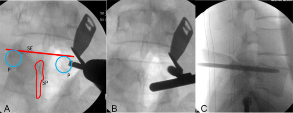
FIGURE 1.

L5 P screw insertion—a perfect PA image of the L5 vertebra is made when the SE is flat to the coronal plane and the SP sits in the midline between the 2 P. The Ps are equidistant from the SP, indicating a perfect PA image without any rotation (A). Start by drilling at the 3-o'clock position on the right P or the 9-o'clock position on the left P with a 3.2-mm drill bit. Fluoroscopy is used to place a 6 × 150-mm Schanz pin into the L5 P and vertebral body. This is checked with PA and lateral images during insertion (B, C).
