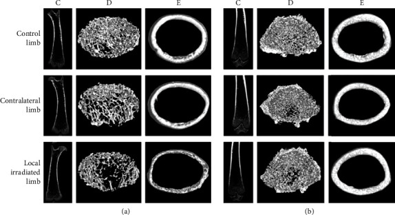
Figure 6.

3D image representation of the femur at 7 and 30 days post single 2 Gy direct irradiation. (a) 7 days and (b) 30 days postirradiation. (A) Coronal cross-sectional femur, (B) 3D femoral trabecular bone, and (C) 3D femoral cortical bone. Image courtesy from Zhai et al. [22].
