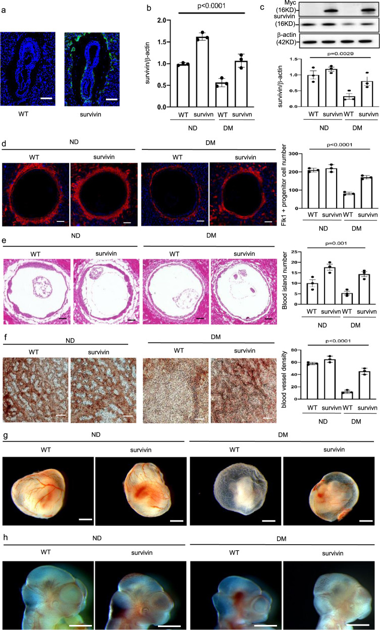
Fig. 5. Survivin overexpression ameliorates maternal diabetes-induced vasculopathy.
a GFP was robustly expressed in Flk-1+ progenitors in the yolk sacs of E8.5 conceptuses. Bars = 200 μm. Survivin mRNA (b) and protein (c) expression in E8.5 embryos. d Imaging and quantification of Flk-1+ progenitors in the yolk sacs of E8.5 conceptuses. Cell nuclei were stained with DAPI (blue). Bars = 100 μm. e Imaging and quantification of blood island numbers in the yolk sacs of E8.5 conceptuses (HE staining). Bars = 100 μm. f Blood vessel density determined by CD31 staining in the yolk sacs of E8.5 conceptuses. Bars = 25 μm. g View of blood vessels in the yolk sacs of E8.5 conceptuses. Bars = 100 μm. h CD31 staining in E8.5 embryos showing disruptive blood vessel formation in embryos exposed to maternal diabetes. Bars = 125 μm. Experiments were performed using three embryos from three different dam per group (n = 3). * indicates significant difference compared to other groups (P < 0.05). ND nondiabetic, DM diabetes mellitus, WT wild type, Survivin Survivin transgenic mice.

