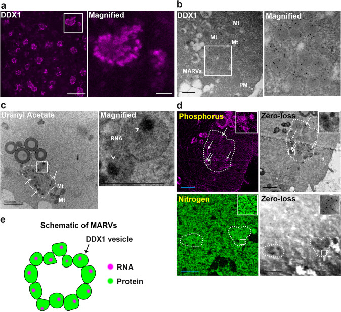
Fig. 1. DDX1 aggregates form ring-like structures with regionalized RNAs in 2-cell embryos.
a STED image of DDX1 staining (magenta) in 2-cell embryos. The white square is magnified in the panel on the right. Scale bar = 5 µm. Magnified image scale bar = 1 µm. Four wild-type crosses were used for each stage (∼8 embryos from each cross). Similar results were obtained for all embryos. b TEM image of immunogold-labelled DDX1 in a 2-cell embryo. An aggregate of DDX1 vesicles (MARV) is shown in the square magnified on the right. Scale bars = 0.5 µm. Four embryos from 2 wild-type crosses were independently collected with similar results obtained for all embryos. c TEM image showing 2-cell embryo stained with OsO4/uranyl acetate. Arrows point to a subset of DDX1 vesicles within MARVs. Six embryos from 3 wild-type crosses were independently collected with similar results obtained for all embryos. Regions containing RNA are indicated by arrowheads. Scale bar = 1 µm. Magnified image scale bar = 0.5 µm. Mt: mitochondria. d Phosphorus (magenta) and nitrogen (green) EFTEM mapping in 2-cell embryos. Scale bars = 1 µm. Zero-loss images are also shown. Four embryos from 2 wild-type crosses were independently collected where similar results obtained for all embryos. A MARV is circled with the dotted line in (c), (d). e Graphical depiction of MARVs.

