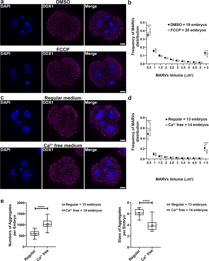
Fig. 3. Sizes of MARVs are linked to mitochondrial potential and Ca2+ distribution/supply.
Two-cell embryos were collected from natural matings. a Embryos were treated with 5 µM FCCP for 2 h. DDX1 aggregates showed reduced sizes in FCCP-treated embryos compared to DMSO control (DDX1, magenta; DAPI, blue). b Statistical analysis of (a) with data from 4 pairs of wild-type natural matings. Data were plotted with Prism. Statistical analysis was performed with two-sided multiple t-test using the Holm-Sidak method, with alpha = 0.05. ****indicates p < 0.0001 (with the exact p < 1E-06). Each circle represents the mean value, and the error bars represent standard deviation. Solid circles represent DMSO-treated control; empty circles represent FCCP-treated. c Embryos cultured in Ca2+ free medium for 24 h show reduced sizes of DDX1 aggregates compared to embryos cultured in regular Ca2+ containing medium. d Size distribution analysis of (c) with data from 3 pairs of wild-type natural matings. Data were plotted with Prism. Statistical analysis was performed with two-sided multiple t-test the Holm-Sidak method, with alpha = 0.05. ****indicates p < 0.0001 (with the exact p < 1E-06 for area <0.5 µm3 and p = 4.5E-05 for area >5 µm3). Each circle represents the mean value, and the error bars represent standard deviation. Solid circles represent regular medium; empty circles represent Ca2+-free medium. e Average aggregate numbers and sizes analysis of (c). Data were plotted with Prism. Statistical analysis was performed with two-sided Students’ t-test. ****indicates p < 0.0001 (with the exact p = 4.26E-06 for left panel and p = 8.29E-07 for right panel). Center line, median; box limits, 25th and 75th percentiles; whiskers, minimum to maximum. Error bars represent standard deviation. Maximum intensity projections of the Z-stack images of each stage are shown in (a), (c). Z-stacks were used for all statistical analyses in this figure. Scale bars = 10 µm.

