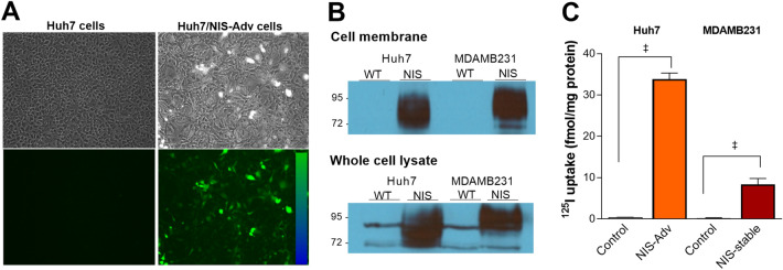
Figure 1.
EGFP and NIS proteins in transiently and stably expressing cancer cells. (A) EGFP fluorescence in Huh7 cells transduced with NIS/EGFP adenovirus (Huh7/NIS-Adv cells). (B) Western blots of NIS protein obtained from the cell membrane fraction (top) and whole cell lysates (bottom) of Huh7, Huh7/NIS-Adv, MDAMB231, and MDAMB/NIS-stable cancer cells. (C) 125I uptake (right) in Huh7/NIS-Adv and MDAMB/NIS-stable cells compared to respective Huh7 and MDAMB control cells. Bars for uptake are mean ± SD of uptake in fmol/mg protein obtained from triplicate samples per group. WT wild type. ‡P < 0.0001.

