
Fig. 4.
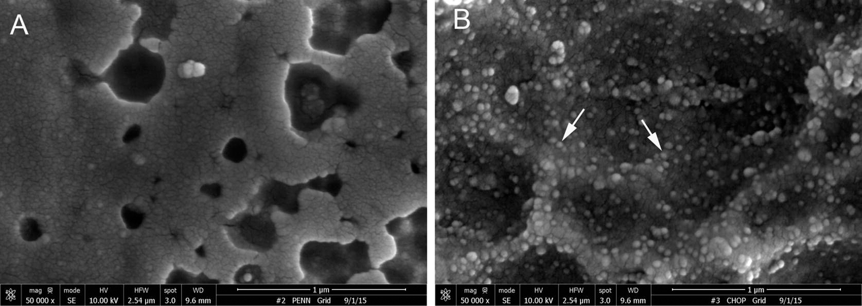
Representative scanning electron microscopy images of stainless steel foil surface modified according to the PABT/PEI(PDT)/protein G/anti-AAV2 antibody protocol AAV2 protocol and either omitted AAV2 incubation (A) or added AAV2 (B). Arrows point to the individual AAV2 particles (B). Original magnification is 80.000×.