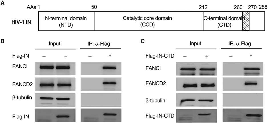
Figure 1. HIV-1 IN interacts with FANCI and FANCD2.

(A) Schematic domain organization of HIV-1 IN.
(B) 293A cells were stably transduced with empty lentiviral vector (−) or lentiviral vector expressing FLAG-tagged full-length HIV-1 IN (+). Cell lysates were treated with benzonase. FLAG-tagged IN was immunoprecipitated by anti-FLAG antibody. The co-immunoprecipitated FANCI and FANCD2 were probed by western blot.
(C) 293A cells were stably transduced with empty lentiviral vector (−) or lentiviral vector expressing flag-tagged CTD of HIV-1 IN (+). Cell lysates were treated with benzonase. FLAG-tagged IN CTD was immunoprecipitated by anti-FLAG antibody. The co-immunoprecipitated FANCI and FANCD2 were probed by western blot.
See also Figure S1.
