FIGURE 1:

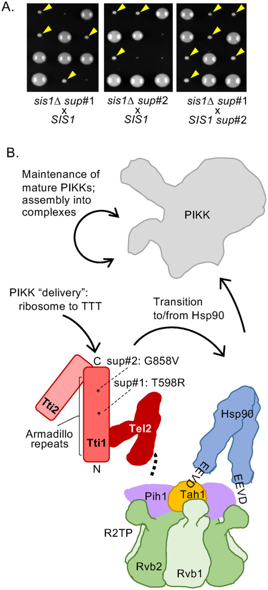
Isolation of spontaneous TTI1 suppressors of cells lacking Sis1. (A) sis1∆ (sis1::LEU2) cells carrying putative suppressor mutations were crossed with a WT SIS1 strain and sporulated and resulting asci dissected onto rich medium. Plates were incubated at 30°C for 4 d (left, suppressor #1 [sup#1]; center, suppressor #2 [sup#2]) and then replica plated to leucine omission media. Colonies that grew (suppressors) are indicated by yellow arrowheads. Right, an sis1∆ sup#1 and a SIS1 sup#2 strain of opposite mating type were crossed and treated as above. All resulting sis1::LEU2 deletion haploids were suppressed for lethality, indicating close linkage of sup#1 and sup#2. (B) Cartoon of TTT complex and possible points of action of Sis1 in PIKK proteostasis (solid arrows). TTT subunits in shades of red, indicating interaction of Tti1 with Tel2 and Tti2. Expanse of α-helical Armadillo Tti1 repeats indicated by bracket; asterisks indicate position of suppressors—sup#1 T598R; sup#2 G858V. Both TTT and Hsp90 have been implicated in folding of PIKKs in coordination with Rvb1/2 in complex with Pih1 and Tah1 as the R2TP complex (or with accessory protein Asa1, not shown). Tel2 interacts physically with the Pih1 subunit (heavy dotted line) and Hsp90 with the Tah1 subunit (via C-terminal EEVD).
